What is Merkel Cell Carcinoma
Merkel cell carcinoma (MCC), also known as neuroendocrine carcinoma of the skin, is a type of skin cancer that occurs when skin cells grow uncontrollably. The tumor usually presents as a single reddish or purplish nodule on a part of the skin that is often exposed to sunlight, such as the face, neck, or arms.
It is a rare disease that affects, in most cases, white individuals over 70 years of age.
Merkel cells are believed to be a type of neuroendocrine cell in the skin because they share some characteristics with nerve cells and hormone-producing cells. They are found mainly at the base of the top layer of the skin, the epidermis, and are very close to the nerve endings of the skin.
Signs and symptoms of Merkel cell carcinoma
Merkel cell carcinoma primarily begins in areas of the skin exposed to the sun, such as the face, neck, and arms, but can occur anywhere on the body. It often first appears as a single shiny pink, red, or purple bump, which usually does not hurt. The skin over the top of the tumor may break open and bleed.
These tumors grow rapidly and can spread as new lumps in nearby skin. They can also reach the lymph nodes, which over time, grow and become visible or are felt as lumps under the skin.
Because they are rare and can resemble other more common types of skin cancer or even non-cancerous skin issues, doctors often do not suspect MCC initially, and the diagnosis is usually made only after a biopsy of the tumor.
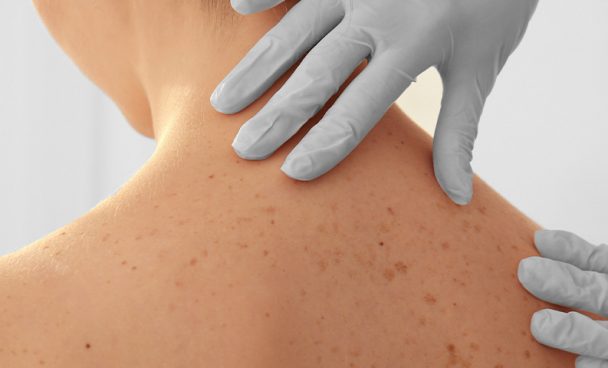
Therefore, it is very important that any new or growing bumps, spots, or lesions be promptly examined by a dermatologist.
Diagnosis of Merkel cell carcinoma
If a patient presents with an abnormal area that may be skin cancer, the doctor will examine it and conduct tests to determine if it is cancer or some other issue. If MCC is diagnosed and there is a risk that it has spread to other parts of your body, further tests will be necessary.
In addition to a standard physical exam, some dermatologists use a technique called dermatoscopy to assess different skin lesions more clearly.
The doctor uses a dermatoscope, which is a special magnifying lens with a light source, used close to the skin.
When there is suspicion of MCC or another type of skin cancer, a small piece of the lesion will be removed and sent to a laboratory for testing – this is called a biopsy.
There are different ways to perform a skin biopsy. The choice is made based on the suspected type of skin cancer, where it is on the body, its size, and other factors. Skin biopsies are done with local anesthesia, and the main types are:
- Tangential biopsy – the upper layers of the skin are scraped off with a small surgical blade. It is useful in diagnosing many types of skin diseases, especially if the doctor considers that an abnormal area should not be a serious skin cancer, such as MCC or melanoma. It is generally not used if the doctor strongly suspects MCC or melanoma because this technique is not deep enough to reach the tumor;
- Punch biopsy – a tool that looks like a small round cookie cutter is used to remove a deeper sample of the skin. The doctor rotates the punch biopsy tool on the skin until it cuts through all necessary layers. The sample is removed, and the edges of the biopsy site are closed;
- Incisional and excisional biopsies – used to examine a tumor that may have grown into deeper layers of the skin. For these types of biopsies, a surgical scalpel is used to cut through the entire thickness of the skin, removing a wedge or piece of skin. The incisional biopsy removes only part of the tumor, while the excisional removes the entire tumor and tends to be preferable for a suspected MCC;
- Lymph node biopsy – MCC can spread to nearby lymph nodes early in the disease. If the tumor has already been diagnosed on the skin, nearby lymph nodes will usually be biopsied to see if the cancer has spread to them. The type of biopsy used depends on the likelihood of the cancer having reached the nearby lymph nodes; and
- Sentinel lymph node biopsy (SLNB) – For Merkel cell carcinoma, we usually use excisional biopsy or less frequently incisional. The lymph node biopsy is restricted to confirmed cases.
If, upon analyzing the samples, the pathologist cannot determine the presence of MCC, special laboratory tests can be performed on the cells to confirm the diagnosis. One of the tests commonly used for MCC is immunohistochemistry (IHC), which looks for certain proteins in the cancer cells, such as CK-20, CK-7, neuro-specific enolase, TTF1, and S100.
If MCC is found, the pathologist will also examine important characteristics, such as the thickness of the tumor, the portion of cells that are actively dividing (mitotic rate), and whether the tumor has invaded the tiny blood vessels or lymph vessels in the sample.
Imaging tests such as magnetic resonance imaging (MRI) or computed tomography (CT) scans to create images of the inside of the body may be performed to check if the MCC has spread to the lymph nodes or to other organs of the body.
These tests may also be done to help check if the treatment is working well or to look for possible signs of cancer coming back (recurrence) after the end of treatment. Understand better the main imaging tests performed in the diagnosis of Merkel cell carcinoma:
- Computed tomography (CT) – can show details in soft tissues (such as internal organs), if lymph nodes are enlarged, or if other organs have suspicious spots (which may be due to the spread of MCC). It also helps guide a needle biopsy;
- Magnetic resonance imaging (MRI) – is very useful in detecting cancer that has spread to the brain and/or spinal cord; and
- Positron emission tomography (PET) – can help show if cancer has spread to the lymph nodes or other parts of the body. It looks for areas where cells are growing rapidly (which may be a sign of cancer), rather than just showing if areas appear abnormal based on their size or shape.
Treatment for Merkel Cell Carcinoma
Surgery is the primary treatment for most Merkel cell carcinomas. Different types can be performed:
- Wide excision – when a diagnosis of MCC is made by skin biopsy, the tumor site will likely need to be surgically cut out (excised) to help ensure that the cancer has been completely removed. This surgery can cure this type of cancer if it has not spread beyond the skin;
- Mohs micrographic surgery – used when the goal is to save as much healthy skin as possible, such as cancer around the eye. Using the Mohs technique, the doctor removes the tumor and a margin of normal-looking skin and examines it under a microscope. If cancer cells are seen at the edges of the removed tissue (the sample), another layer of skin is removed and examined. This is repeated until the skin samples do not contain cancer cells;
- Lymph node dissection – if cancer is found in nearby lymph nodes, a lymph node dissection is usually performed. In this operation, the surgeon removes all lymph nodes near the primary tumor – for example, if Merkel cells are found in an arm, the surgeon will remove the lymph nodes from the armpits (axillary) on that side of the body, as these nodes are where cancer cells would be most likely to arrive first; and
- Skin grafting and reconstructive surgery – after removing large skin cancers, it may not be feasible to stretch the skin close enough to sew the edges of the wound and cover the entire area. In these cases, healthy skin can be taken from another part of the body and grafted onto the exposed area to aid in healing and improve appearance.
There is no consensus on how radiotherapy should be used for Merkel cell carcinoma, but it is known that it can be used in the following situations:
- To treat the area of the primary skin tumor (primary) after surgery, trying to kill any cancer cells that may have been left behind;
- To treat the primary tumor if surgery is not an option (due to the person not being healthy enough or the tumor being in a place where it cannot be fully removed);
- To treat the lymph nodes near the primary tumor;
- To help treat MCC that has returned after surgery, either in the skin or in the lymph nodes; and
- To help treat MCC that has spread to distant parts of the body, usually along with other treatments.
These days, standard systemic therapy is immunotherapy. This therapy is approved and available for those with unresectable (unable to be removed by surgery) or metastatic disease (when it has already spread to other organs).
Prevention of Merkel Cell Carcinoma
Since Merkel cell carcinoma can be caused by sun exposure, some strategies to prevent it are important:
- Avoid excessive sun exposure between 10 a.m. and 4 p.m. when the sun’s rays are strongest;
- Do not use artificial tanning beds;
- Wear protective clothing, such as long-sleeved shirts, pants, and a wide-brimmed hat, especially when you need to be exposed to the sun between 10 a.m. and 4 p.m.; and
- Use sunscreen daily, with a minimum sun protection factor (SPF) of 30 and protection against UVA and UVB rays, reapplying every two hours and/or after swimming or sweating.
Other known, uncontrollable risk factors include:
- Being over 50 years old;
- Having fair skin; and
- Having a compromised immune system, which includes people with HIV, multiple myeloma, melanoma, or chronic leukemia and people taking immunosuppressive medications.
