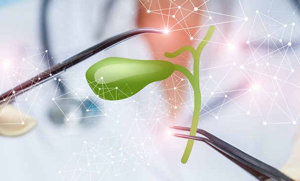
What is gallbladder cancer?
Gallbladder cancer usually does not cause signs or symptoms until later in the course of the disease, when the tumor is large. If the cancer is detected at an earlier stage, treatment has a better chance of success. To understand this cancer, it’s helpful to know about the gallbladder and how it functions.
The gallbladder stores and concentrates bile, a fluid produced in the liver that helps digest fats in food as they pass through the small intestine. When food is being digested, the gallbladder releases bile through a small tube called the cystic duct.
The gallbladder is a small pouch located just below the liver. Bile aids in fat digestion, but the gallbladder itself is not essential. Its removal in a healthy individual typically does not cause health or digestion problems.
Subtypes of gallbladder cancer
Gallbladder cancers are rare, and almost all are adenocarcinomas, but there are other types that are even less common. The subtypes of this disease, therefore, are:
-
- Adenocarcinoma – begins in gland-like cells that line many surfaces of the body, including the inside of the digestive system;
- Papillary adenocarcinoma – also called papillary cancer, is a rare type of gallbladder adenocarcinoma, with a better prognosis than most other types of gallbladder adenocarcinomas. The cells in these cancers are organized into finger-like projections. Generally, papillary cancers are less likely to spread to the liver or nearby lymph nodes;
- Adenosquamous carcinoma;
- Squamous cell carcinoma;
- Carcinosarcomas.
Symptoms and signs of gallbladder cancer
The signs and symptoms of gallbladder cancer usually only manifest when the disease is already in an advanced stage, but in some cases, they can appear at an earlier stage when treatment is more effective. Some of the most common symptoms of gallbladder cancer are:
- Abdominal pain (typically in the upper right abdomen);
- Nausea and vomiting;
- Jaundice (yellowing of the skin and whites of the eyes);
- Abdominal lumps.
Other less common symptoms include:
- Loss of appetite;
- Unexplained weight loss;
- Fever;
- Severe itching;
- Pale stools;
- Dark urine.
Diagnosis of gallbladder cancer
Some gallbladder cancers are found after the gallbladder is removed because of gallstones or to treat chronic inflammation. However, most gallbladder cancers are not detected until a person seeks help because of symptoms.
During a clinical consultation, the doctor will examine the abdomen, check the color of the skin and eyes, and look for swelling in the lymph nodes. If the symptoms and/or physical exam suggest gallbladder cancer, tests will be ordered. This may include laboratory tests, imaging tests, and other procedures.
Here are the main ones:
- Liver and gallbladder function tests – laboratory tests may be performed to determine the amount of bilirubin in the blood, as problems in the gallbladder, bile ducts, or liver can elevate the level of this substance. Tests for albumin, liver enzymes (alkaline phosphatase, AST, ALT, and GGT), and other specific substances may also be requested.
- Tumor markers – are substances produced by cancer cells that can be found in the blood. People with gallbladder cancer may have elevated levels of markers called CEA and CA 19-9 in the blood. These markers are not specific to gallbladder cancer – meaning other types of cancer or even some other health conditions can also increase them.
- Imaging tests – such as computed tomography (CT) scans, magnetic resonance imaging (MRI), or ultrasound – may be done for a variety of reasons: to look for areas suspicious for cancer, to help guide a needle for sampling and biopsy, to see if and how far the cancer has spread, and to determine the best treatment.
- Ultrasound – is usually the first imaging test done in people with symptoms such as jaundice or pain in the upper right abdomen. It is easy to perform and does not use radiation. It can also be used to guide a needle into the suspicious area or lymph node so that cells can be removed and examined under a microscope. This is called an ultrasound-guided needle biopsy.
- Endoscopic or laparoscopic ultrasound – the doctor places the ultrasound transducer inside the body and near the gallbladder – this provides more detailed images than a standard ultrasound. The transducer is at the end of a thin, lighted tube that contains a camera; the tube is passed through the mouth, through the stomach, and near the gallbladder (endoscopic ultrasound) or through a small surgical cut in the belly (laparoscopic ultrasound). The exam helps to see if and how far the cancer may have spread into the wall of the gallbladder.
- Computed tomography (CT) – can be used to help diagnose gallbladder cancer, showing tumors in the area, and is also useful in staging. It can show organs near the gallbladder (especially the liver), as well as lymph nodes and distant organs to which the cancer may have spread.
CT angiography can be used to observe blood vessels near the gallbladder, helping to confirm if surgery is an option. In biopsy, CT helps guide the needle to the tumor mass to remove part of the tissue for analysis.
- Magnetic resonance imaging (MRI) – shows detailed images of soft tissues in the body. A contrast material called gadolinium can be injected into a vein before the exam to see the details even better.
- Cholangiography – an imaging test that examines the bile ducts to see if they are blocked, narrowed, or dilated (enlarged). A tumor may be blocking a duct. The test also helps in planning surgery.
- Angiography – an X-ray test used to observe blood vessels. A thin plastic tube (catheter) is inserted into an artery, and a small amount of contrast is injected to outline the blood vessels. Then, X-rays are taken. The images show if blood flow in an area is blocked or affected by a tumor, as well as any abnormal blood vessels in the area, helping to plan surgery. Angiography can also be done with a computed tomography (CT) scan (CT angiography) or a magnetic resonance imaging (MRI) scan (MR angiography).
- Laparoscopy – the doctor inserts a thin tube with a light and a small video camera on the end (laparoscope) through a small incision (cut) in the front of the abdomen to view the gallbladder, liver, and other nearby organs and tissues. If needed, doctors can also insert special instruments through the incisions to take biopsy samples for testing.
- Biopsy – during a biopsy, the doctor removes a sample of tissue to be examined under a microscope for cancer cells. In most types of cancer, a biopsy is necessary to confirm the diagnosis, but it is not always done before surgery to remove a gallbladder tumor because there is a risk that manipulating the tumor with the needle could cause the cancer cells to spread.
If imaging tests show a tumor in the gallbladder and there are no clear signs that it has spread, the doctor may decide to proceed directly to surgery and treat the tumor as gallbladder cancer.
Treatment of gallbladder cancer
The main treatment is surgical. There are two general types of surgery for gallbladder cancer:
- Potentially curative surgery (resectable disease) – performed when imaging tests or the results of previous surgeries show that there is a good chance the surgeon can remove all the cancer.
- Palliative surgery – performed to relieve pain symptoms or treat (or even prevent) complications, such as bile duct blockage. This type of surgery is done when the tumor is too widespread to be completely removed and does not cure cancer, but it can help the patient feel better and live longer.
In addition to surgery, radiation therapy can be used, usually after surgery, to kill any cancer cells left behind (adjuvant therapy), or as part of the treatment in advanced cancers that have spread widely throughout the body and cannot be removed. It is also used as palliative therapy, relieving symptoms of very advanced cancer.
Some side effects of radiation therapy are:
- Skin problems resembling sunburns (redness, blisters, and peeling in the treated area);
- Nausea and vomiting;
- Diarrhea;
- Fatigue;
- Liver damage.
Chemotherapy is another type of treatment for gallbladder cancer. It can be oral or intravenous, and it can be given with the goal of curing the patient (after surgery with complete removal of the tumor) or just to control the disease (when complete surgical removal of the tumor is not possible). Rarely, chemotherapy can be given together with radiation therapy to increase its effectiveness.
Prevention of Gallbladder Cancer
Knowing the risk factors for developing gallbladder cancer can be a way to prevent it – at least in relation to avoidable factors, such as smoking. Factors that cannot be changed, such as family history or presence of a specific health condition, should be monitored to minimize the risk of diagnosing cancer only at an advanced stage.
Regarding gallbladder cancer, its development may be related in some way to chronic inflammation (long-term irritation and swelling) in the gallbladder.
Other risk factors include:
- Gallstones – are the most common risk factor for this cancer. Gallstones are collections of cholesterol and other substances that form in the gallbladder and can cause chronic inflammation. Up to four out of five people with gallbladder cancer have gallstones when they are diagnosed;
- Porcelain gallbladder – a condition in which the wall of the gallbladder is covered with calcium deposits. It sometimes occurs after long-term inflammation of the gallbladder (cholecystitis), which can be caused by gallstones. People with this condition have a higher risk of developing gallbladder cancer, possibly because the two conditions may be related to inflammation;
- Sex – gallbladder cancer occurs more in women than in men. Gallstones and inflammation of the gallbladder are important risk factors for gallbladder cancer and are also much more common in women than in men;
- Obesity – patients with gallbladder cancer are more often obese or overweight than people without this disease. Obesity is also a risk factor for gallstones, which may help explain this link;
- Age – gallbladder cancer is seen mainly in older people, but younger people can also develop it. The average age of people when they are diagnosed is 72;
- Gallbladder polyps – are a “nodule” that projects into the surface of the gallbladder’s inner wall. Some polyps are formed by deposits of cholesterol in the gallbladder wall, others may be small tumors (cancerous or not) or may be caused by inflammation. Polyps larger than 1 centimeter are more likely to be cancerous. Therefore, doctors often recommend gallbladder removal in patients with polyps of this size or larger;
- Smoking;
- Exposure to chemicals used in rubber and textile industries;
- Exposure to nitrosamines.
